Lumbar Microscopic Discectomy

Lumbar Microscopic Discectomy

Lumbar Microscopic Discectomy is a specialized surgical procedure designed to treat lumbar disc herniations, which occur in the lower back. This minimally invasive technique aims to relieve pressure on the spinal nerves or spinal cord caused by a herniated or bulging disc. The procedure involves the use of a high-powered microscope, which provides the surgeon with a magnified view of the affected area, allowing for precise and accurate removal of the problematic disc material.

Procedure Overview

The surgery typically begins with the patient under general anesthesia. The surgeon makes a small incision in the lower back to access the lumbar spine. Using the microscope, the surgeon can see the spinal structures in great detail, which enables the careful removal of the herniated disc material that is pressing on the nerves or spinal cord. The small incision minimizes disruption to surrounding tissues, reducing postoperative pain and promoting quicker recovery.

During the procedure, the surgeon carefully navigates through the muscles and other tissues to reach the affected disc. The microscope allows for enhanced visualization, which is crucial for removing only the necessary portion of the disc without damaging the surrounding structures. This precision helps to preserve as much healthy tissue as possible, further aiding in recovery and reducing the risk of complications.

What is a Lumbar Microscopic Discectomy?

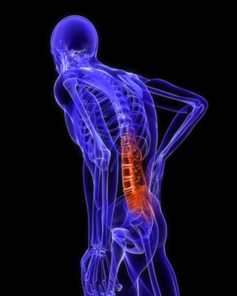
Lumbar microscopic discectomy is a surgical procedure used to treat lumbar disc herniations in the lower back. This minimally invasive technique aims to relieve pressure on the spinal nerves or spinal cord caused by a herniated or bulging disc. Here’s a detailed overview:

Meaning

Lumbar Microscopic Discectomy is a specialized form of discectomy that utilizes a microscope to enhance the surgeon’s view of the spinal structures. The microscope provides magnified, detailed visualization of the affected disc and surrounding tissues, allowing for precise and targeted removal of the herniated disc material. This technique contrasts with traditional open discectomy, which involves larger incisions and more extensive tissue disruption.

How It Works

Pre-Surgery Preparation: The procedure typically starts with the patient under general anesthesia. Preoperative imaging, such as MRI or CT scans, helps in identifying the exact location and extent of the disc herniation.

Incision: The surgeon makes a small incision in the lower back to access the lumbar spine. The size of the incision is significantly smaller compared to traditional open surgery, which reduces tissue damage and accelerates recovery.

Microscopic Visualization: A high-powered microscope is used to magnify the surgical site. This detailed view allows the surgeon to navigate through the spinal structures more accurately and to precisely target the herniated portion of the disc.

Disc Material Removal: Through the small incision, the surgeon removes the part of the herniated disc that is pressing on the nerves or spinal cord. The use of the microscope ensures that only the problematic disc material is removed while preserving as much healthy tissue as possible.

Closure and Recovery: After the herniated disc material is removed, the incision is closed with sutures or staples. The minimally invasive nature of the procedure usually leads to less postoperative pain and a quicker recovery compared to traditional discectomy.

What are the Indications

Lumbar microscopic discectomy is often recommended for patients who experience significant symptoms from a herniated disc that have not improved with conservative treatments like physical therapy, medication, or steroid injections. Symptoms indicating the need for surgery may include persistent lower back pain, sciatica (pain radiating down the leg), and neurological deficits such as numbness or weakness in the legs.

Recovery

Post-surgery, patients usually need a period of rest and may benefit from physical therapy to strengthen the back muscles and improve flexibility. Recovery times vary, but many patients can resume light activities within a few weeks and return to their normal activities within a few months.

In summary, lumbar microscopic discectomy is an effective, minimally invasive option for addressing lumbar disc herniations, offering a promising approach for those seeking relief from severe back pain and associated symptoms.

Types of Lumbar Microscopic Discectomy

Lumbar Microscopic Discectomy, a minimally invasive surgical procedure to remove herniated disc material in the lower back, can be performed using various techniques and approaches. Here are the primary types or variations of lumbar microscopic discectomy:

Standard Microscopic Discectomy

  • Procedure: This is the most common type, where the surgeon uses a microscope to enhance visualization of the herniated disc and surrounding structures. A small incision is made in the lower back, and the microscope allows precise removal of the herniated disc material.
  • Advantages: The use of the microscope improves accuracy, minimizes tissue damage, and reduces recovery time compared to traditional open surgery.
  • Microendoscopic Discectomy
  • Procedure: This technique combines endoscopic technology with microscopic visualization. An endoscope, a thin tube with a camera and light, is used alongside the microscope to provide enhanced views of the surgical site through a small incision.
  • Advantages: This approach offers even greater precision, less tissue disruption, and potentially faster recovery due to the minimally invasive nature of the endoscope.

Percutaneous Endoscopic Lumbar Discectomy (PELD)

  • Procedure: In this procedure, an endoscope is inserted through a small incision to remove the herniated disc material. The surgeon uses real-time imaging and the endoscope to navigate to the affected area.
  • Advantages: PELD is highly minimally invasive, resulting in minimal scarring, reduced pain, and quicker recovery. It is particularly useful for specific types of herniations and patients with certain anatomical considerations.

Transforminal Lumbar Interbody Fusion (TLIF)

  • Procedure: Although TLIF is primarily a fusion procedure, it can be combined with a microscopic discectomy. The surgeon accesses the spine from the side of the vertebra, removing the herniated disc material and then placing a bone graft to promote fusion.
  • Advantages: This approach is beneficial for patients who require stabilization of the spine in addition to the removal of herniated disc material.

Minimally Invasive Discectomy

  • Procedure: This general category includes various techniques that employ smaller incisions and specialized instruments to remove herniated disc material. The goal is to achieve the same results as traditional discectomy with less tissue damage and quicker recovery.
  • Advantages: These procedures typically involve less postoperative pain, shorter hospital stays, and faster return to normal activities.

Tubular Discectomy

  • Procedure: A tubular retractor system is used to create a small working channel through which the surgeon can perform the discectomy using microscopic visualization. The tube helps to minimize tissue disruption and maintain stability.
  • Advantages: This technique allows for precise removal of herniated disc material while preserving surrounding muscles and tissues, leading to quicker recovery.

Lumbar Microscopic Discectomy

Symptoms of Lumbar Microscopic Discectomy

Lumbar microscopic discectomy is a minimally invasive surgical procedure used to treat herniated discs in the lower back (lumbar spine). The symptoms that typically lead to considering this procedure include:

  • Radiating Pain: Pain that radiates from the lower back down one leg, commonly known as sciatica. This pain may be sharp, shooting, or burning and can worsen with movement, coughing, or sneezing.
  • Lower Back Pain: Persistent or acute pain localized to the lower back, often exacerbated by certain activities such as bending, lifting, or prolonged sitting or standing.
  • Numbness and Tingling: Sensations of numbness, tingling, or “pins and needles” in the buttock, leg, or foot. These symptoms may indicate compression or irritation of spinal nerves by the herniated disc.
  • Muscle Weakness: Weakness in the leg or foot muscles, making it difficult to lift the affected limb, walk normally, or perform daily activities that require lower body strength.
  • Loss of Bowel or Bladder Control: In rare cases, severe compression of nerves in the lumbar spine can lead to loss of control over bowel or bladder function. This is a medical emergency and requires immediate attention.

Purpose of Lumbar Microscopic Discectomy:

The procedure aims to relieve pressure on the spinal nerves caused by the herniated disc, thereby alleviating symptoms and promoting healing. During the surgery:

  • Minimally Invasive Approach: Small incisions are made near the spine, through which specialized instruments and a microscope are used to remove the portion of the herniated disc pressing on the nerve.

  • Preservation of Surrounding Tissues: Unlike traditional open surgery, microscopic discectomy minimizes disruption to muscles, ligaments, and other soft tissues, leading to faster recovery times and reduced postoperative pain.

Recovery and Rehabilitation:

Following surgery, patients typically experience:

  • Pain Relief: Immediate relief from leg pain and gradual reduction in lower back pain.

  • Gradual Return to Activities: Patients are encouraged to resume light activities soon after surgery and gradually increase activity levels under the guidance of healthcare providers.

  • Physical Therapy: Rehabilitation exercises to strengthen the back muscles, improve flexibility, and prevent future spine problems.

Considerations:

While lumbar microscopic discectomy is generally safe and effective, it may not be suitable for all patients or types of herniated discs. Factors such as the location and size of the herniation, overall health, and response to conservative treatments influence the decision for surgery.

In conclusion, lumbar microscopic discectomy is a valuable option for relieving symptoms of herniated discs in the lower back, particularly when conservative treatments have not provided sufficient relief. By targeting the source of nerve compression with minimal tissue disruption, this procedure aims to restore function and quality of life for individuals experiencing debilitating pain and neurological symptoms due to lumbar disc herniation.

Conclusion 

Lumbar microscopic discectomy is a highly effective, minimally invasive procedure designed to alleviate pain and neurological symptoms caused by herniated discs in the lower back. While recovery typically involves some common postoperative symptoms such as pain, soreness, and fatigue, these generally diminish over time with proper care and rehabilitation. The procedure’s minimally invasive nature often results in faster recovery, less postoperative pain, and a quicker return to normal activities compared to traditional open surgery.

For those seeking expert care in Indore, Dr. Amit Deora, a renowned neurosurgeon in Indore, offers specialized expertise in lumbar microscopic discectomy and other spinal treatments. Dr. Deora’s advanced surgical techniques and personalized approach ensure that patients receive the highest quality of care tailored to their specific needs. Consulting with Dr. Amit Deora can provide you with a comprehensive evaluation and effective treatment options to help you achieve optimal recovery and an improved quality of life.